In addition to examinations and various treatments, our Gynecologic Center also offers an outstanding number of surgical cases. Since the opening of the institution, our team has performed more than 1,000 successful gynecological surgeries, with a total of more than 400 in 2023 alone. Last year, endometriosis and myoma were the most prominent cases.
This is also important because, in addition to experienced specialists, the well-accustomed operating theatre also plays a key role in the success of a possible operation, and this kind of safety and experience works well in the institution. The patient’s journey is assisted and coordinated by the case manager. This person supports the patient from the first specialist examination to the recovery phase after the operation and is responsible for ensuring that there are no open questions and that the patient is aware of the processes, tasks, and appointments with the least follow-up and workload.
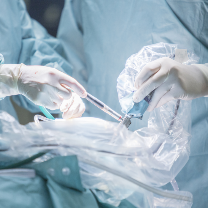
The surgeon controls a robotic system to perform the procedure. The doctor makes a few small incisions and uses a 3D HD camera to get a crystal-clear, magnified view of the uterus, cervix, and surrounding tissues. The surgeon sits next to you at a console (surgical console) and operates through the incisions with small instruments and a camera. The da Vinci surgical system maps the surgeon’s hand movements in real time by bending and rotating instruments similar to the human hand, but with a greater range of motion.
During the procedure, the doctor enters the abdominal cavity through a small, buttonhole-sized opening on the abdomen. An optic equipped with a camera system is introduced through the incision made at the navel, and the resulting image can be viewed on a high-resolution monitor. Because of orientation in the abdominal cavity, the abdomen is filled with CO2 (carbon dioxide) gas, so the abdominal organs become clearly visible. The surgeon does not insert his hand into the abdominal cavity, but uses long, thin medical device (manipulator) instead. The procedure is very safe and not stressful for the patient. This type of surgery is used for the removal of myomas, for the solution of formulas in the ovaries, and during hysterectomy. In fact, certain surgical oncology procedures (extended hysterectomy and pelvic lymph node removal) are also performed with this procedure. Preserving the integrity of the abdominal wall ensures that the patient can go home the next day and live his normal life.
In this surgical procedure, we penetrate the uterine cavity from the vagina, so the abdominal cavity remains unaffected. A thin device is inserted through the cervical canal of the uterus, which includes a camera, a manipulator working channel and a suction system for introducing a fluid. We also see the structure of the uterine cavity on a high-resolution monitor. This procedure is used to resolve abnormalities in the uterine cavity (polyp, septum, myoma distorting the uterine cavity). The method requires little effort, is easy to perform, and the patient can leave the institution a few hours after the operation.
During the procedure, we enter the abdominal cavity from the vagina and perform interventions through the vagina. Vaginal prolapse, urinary or stool retention disorders can be treated with this surgical procedure. This method also involves very little strain on the patient and is an aesthetically pleasing operation.