Chest CT and lung CT scans are used to examine the chest organs, lungs, chest vessels, surrounding lymph nodes, the chest section of the trachea and oesophagus, and the diaphragm.
In some cases, the tests can be done as a screening, and you can also request a lung screening on your own, so for information about chest X-rays and low-dose lung CT scans, see the “lung screening link”.
Chest CT scans are only performed with or without the use of contrast agents on the advice of a doctor, depending on the questions asked by your specialist.
CT scans are done with X-rays so they are not used in pregnancy.

Chest CT, lung CT scans can be performed without the use of contrast agents natively, to diagnose inflammation, pulmonary fibrosis or bone fractures.
However, chest CT scans are predominantly complete with the administration of contrast material to form an opinion. The search for various tumours, tumours, lesions, metastases following an existing tumour, lymph nodes and their controls are also sufficiently informative as contrast material examinations.
For the examination of the thoracic large vessels, aorta, arteries, angiographic contrast studies with vascular staining are recommended.
For the examination of pulmonary arteries, a special pulmonary CT angiography is performed in case of suspected pulmonary embolism. Due to the acute nature of the investigation, these are mainly performed in cases requiring immediate attention. In many cases, suspicion may be suggested by an elevated value of a laboratory parameter called D-dimer, so any investigations that arise because of this should also be done urgently.

Contrast material allows for more detailed imaging, and some details can only be detected with contrast material. However, it is not recommended in cases of known iodine contrast agent allergy or renal stenosis because of the health risk. For this reason, a renal function lab within two weeks before the chest CT will be required.
The results of the renal function lab will be used to consider the use of contrast. No contrast will be used below an eGFR of 45 due to the inherent risks.
Contrast media may interact adversely with metformin, a commonly used antidiabetic drug used to treat diabetes. Therefore, you should stop taking metformin-containing medicines for two days before and after the test. Be sure to follow your doctor’s instructions.
In the case of known thyroid problems, including hyperthyroidism, consult your doctor about contraindications to contrast media.
It is important not to eat for 4-6 hours before using contrast media, but you can drink water.
A chest CT scan is a non-invasive, painless test, you may only feel discomfort when the needle is inserted to give the contrast agent or from its effects (warmth, hotness, urination, bowel movements).
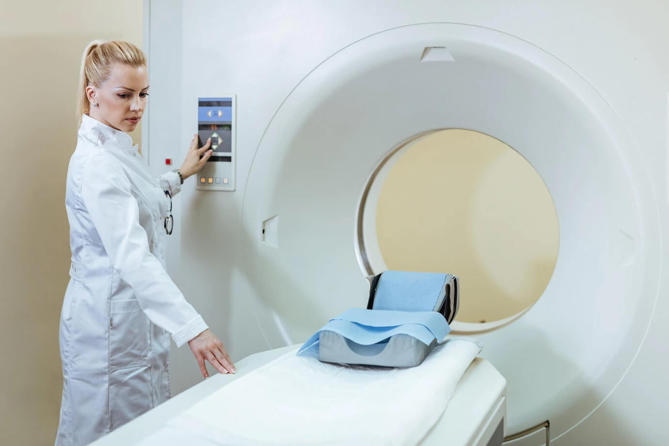
During the examination you will be asked to lie on the moving table of the CT machine. During the scan, you will be instructed to take in air at times, so you will need to hold your breath for 20-30 seconds while the machine takes a series of images.
Scans without contrast material take less time, but apart from the time it takes to secure the vein and inject the material, they are only a few minutes longer than those with contrast material.
Make an appointment for a lung CT scan with the excellent specialists at Wáberer Medical Center, or call us at +36-1-323-7000 to have your scan done at our well-equipped clinic in Buda without a long wait.